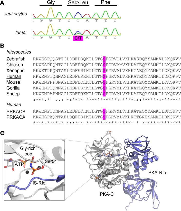
Figure 1. Somatic mutation at the conserved serine 54 of PRKACB.
(A) Partial electropherograms of the PRKACB gene showing a canonical sequence in the leukocytes of the patient and the c.161C>T (S54L) mutation in the tumor (according to the variant NM_002731). (B) Multiple alignments of PRKACB sequences (performed with CLUSTAL O 1.2.1) showing that the residue S54L is conserved between different isoforms and species. (C) Crystal structure of the PKA holoenzyme complex. The catalytic subunit (Cα) is in white, and the regulatory subunit (RIα) is in blue. On the left, magnification of the active site shows Ser54 of the glycine-rich loop, which is involved in binding of the cosubstrate ATP (orange sticks) and substrates (here the pseudosubstrate sequence of RIα). S54L is located in the immediate vicinity of the inhibitor sequence of the regulatory subunit. The structure (PDB 2QCS) was visualized using PyMOL v1.3 (Schrödinger LLC).

