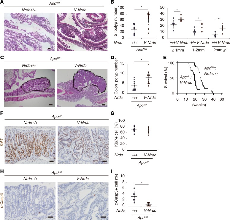
Figure 3. Forced expression of NRDC in epithelial cells enhances ApcMin mouse intestinal tumors.
(A) Representative H&E staining of the small intestines of ApcMin and ApcMin; Villin-Nrdc (V-Nrdc) mice. (B) ApcMin; Villin-Nrdc mice showed a greater increase in tumor formation in the small intestine (SI) than ApcMin mice (n = 6–11). *P < 0.05 by unpaired 2-tailed Student’s t test. Total number (left) and number in each size fraction (right) are depicted. (C) Representative H&E staining of colon tumors (dotted area) in ApcMin and ApcMin; Villin-Nrdc mice. (D) The numbers of colon tumors in ApcMin and ApcMin; Villin-Nrdc mice (n = 11). *P < 0.05 by unpaired 2-tailed Student’s t test. (E) A Kaplan-Meier analysis demonstrated that ApcMin; Villin-Nrdc mice showed a significantly shorter survival compared with ApcMin mice (n = 11). *P < 0.001 by log-rank test. (F) Immunostaining for Ki67 in ApcMin and ApcMin; Villin-Nrdc mice. (G) There was no difference in Ki67-positive cells between ApcMin and ApcMin; Villin-Nrdc mice (n = 7). (H) Immunostaining for cleaved caspase-3 in ApcMin and ApcMin; Villin-Nrdc mice. (I) The percentage of cleaved caspase-3–positive apoptotic cells was decreased in ApcMin; Villin-Nrdc mice (n = 7). *P < 0.05 by unpaired 2-tailed Student’s t test. All scale bars: 100 μm.

