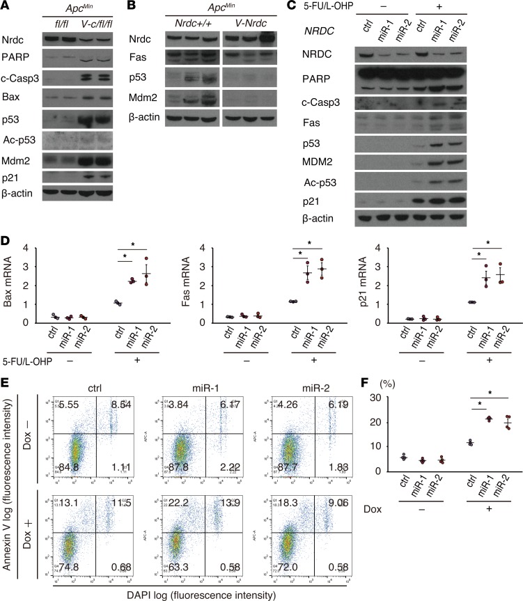
Figure 4. NRDC regulates apoptosis and chemosensitivity of HCT116 cells.
(A) Western blotting of small intestine lysates from ApcMin and ApcMin; Villin-Nrdc mice was performed with the indicated antibodies. (B) Western blotting of small intestine lysates from ApcMin; Nrdcfl/fl and ApcMin; Villin-cre; Nrdcfl/fl mice. Fas, p53, and Mdm2 were decreased in ApcMin; Villin-Nrdc mice. (C) HCT116 cells, in which NRDC was knocked down (control, miR-1, miR-2), were treated with or without 5-fluorouracil (5-FU, 5 μM) and oxaliplatin (L-OHP, 1 μM) for 48 hours and harvested for Western blotting with the indicated antibodies. (D) qRT-PCR analysis showed that Bax, Fas, and p21 mRNA levels were increased by NRDC knockdown. All data represent means ± standard error (SE) of 3 independent experiments with duplicate samples. (E) Representative images of flow cytometric analysis by annexin V and DAPI staining in HCT116 cells. Cells with or without NRDC knockdown were analyzed after the treatment with doxorubicin (Dox) for 24 hours. (F) Quantification of apoptotic cells (annexin V-positive and DAPI-negative) (Q1). All data represent means ± SE of 4 independent experiments with duplicate samples. *P < 0.05 by 1-way ANOVA with Tukey-Kramer post hoc test.

