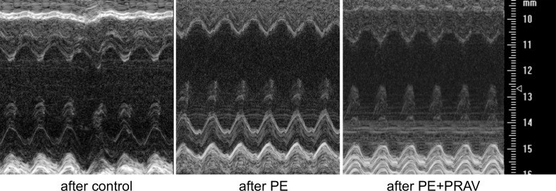
Figure 4. Transthoracic views of the maternal hearts 60 days after pregnancy, acquired using Vevo770 ultrasound system.
Representative M-mode images of the left ventricle (LV) of maternal hearts after a normal pregnancy (control mice), preeclamptic pregnancy (PE mice), and preeclamptic pregnancy treated with pravastatin (PE+PRAV). The y axis represents the distance (mm) from the transducer; time is shown on the x axis. The M-mode images show the LV anterior wall, LV chamber, and LV posterior wall throughout diastole and systole. Echogenic peaks visible along the PW during systole represent the papillary muscle entering the field of view. Images were acquired using the Vevo 770 ultrasound system (VisualSonics).

