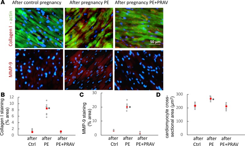
Figure 5. Representative microphotographs of the left ventricle in maternal hearts after a normal pregnancy and pregnancies complicated by preeclampsia (with and without pravastatin treatment).
(A) Increased collagen deposition (red fluorescence, top panels), indicative of fibrosis, and MMP-9 (red fluorescence, bottom panels), indicative of tissue remodeling, are observed in the maternal heart 60 days after preeclampsia. Green fluorescence in top panels indicates F-actin within the cardiomyocytes. Pravastatin prevented LV fibrosis/remodeling. Ten slides per experimental group (5–7 mice/experimental group) were analyzed. Scale bar: 50 μm. (B and C) Quantification of staining for collagen I and MMP-9 expressed as a percentage of the tissue area. Ten slides per experimental group were analyzed for collagen content and MMP-9 expression. Raw data and mean ± SD are shown. Comparisons between groups were performed by 1-way ANOVA with Bonferroni’s post hoc test. *P < 0.01, different from control. (D) Quantification of cross-sectional area of cardiomyocytes in left ventricles. Red squares represent the mean. Areas of 50 myocytes from 5 randomly selected microscope fields from the LV posterior wall from each experimental group (n = 5–7 mice/experimental group) were averaged to represent the myocyte area. *P < 0.05, different from control.

