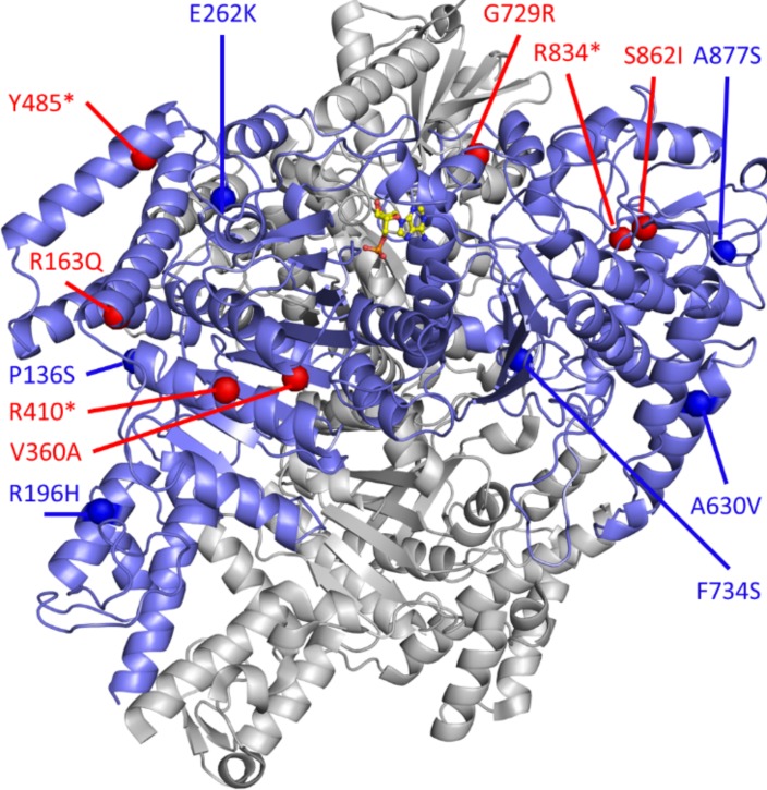
Figure 9. 3-D protein structure of DHTKD1/OGDHL.
Dehydrogenase E1 and transketolase domain–containing 1 (DHTKD1)/oxoglutarate dehydrogenase-like (OGDHL) (blue) complexed with AMP (silver); the structure is built based on a homology model of E. coli sucA. The residue position of missense variants and starting point of frameshifts or truncations are indicated for DHTKD1 (red spheres) and OGDHL (blue spheres).

