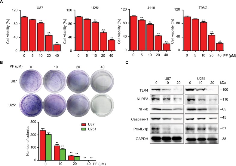
Figure 1.
The effects of paeoniflorin on cell proliferation and TLR4 signaling in glioblastoma cells.
Notes: (A) U87, U251, U118, and T98G cells were treated with different concentrations of paeoniflorin for 24 h and after which cell viability was examined using the CCK-8 assay. (B) U87 and U251 cells were cultured for 7 days after treatment with varying concentration of paeoniflorin and stained with 0.1% crystal violet. Colonies containing >50 cells were counted (C) in U87 and U251 cells that were incubated with the indicated concentrations of paeoniflorin for 24 h; Western blotting was then performed to analyze TLR4 protein expression levels as well as downstream effectors: n=3 and n=4 for cell counts and Western blotting, respectively. All tests were performed in triplicate. **P<0.01 compared with control (0 μM).
Abbreviations: GAPDH, glyceraldehyde-3-phosphate dehydrogenase; IL, interleukin; NF-κB, nuclear factor κB; NLRP, nucleotide-binding domain and leucine-rich repeat containing protein 3; CCK-8, Cell Counting Kit-8; TLR4, Toll-like receptor 4; PF, paeoniflorin.

