Figure 3.
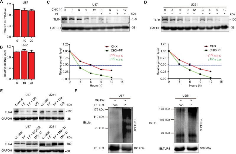
Paeoniflorin promotes TLR4 degradation via the ubiquitin–proteasome pathway in glioblastoma cells.
Notes: (A, B) TLR4 mRNA expression was detected by quantitative real-time polymerase chain reaction in cells treated with different concentrations of paeoniflorin and was normalized to GAPDH expression. Expression was expressed as a fold change relative to 0 μM paeoniflorin-treated U87 and U251 cells. (C, D) Time course of TLR4 degradation. Top panel, CHX (100 μg/mL) was added to U87 and U251 cells treated with or without 20 μM paeoniflorin for 24 h, after which Western blot analysis was performed. Bottom panel, quantified TLR4 band intensities, which are representative of 3 separate analyses by Image J. The relative intensities of each band from the cell samples were quantified by densitometry as a function of time, with the dotted line (---) indicating the half-life (T½) of TLR4 protein in U87 and U251 cells. (E) U87 and U251 cells were incubated with 20 μM CQ or 5 μM MG-132 for 6 h before being treated with 20 μM paeoniflorin or PBS for 18 h. TLR4 protein expression was estimated by Western blotting. (F) U87 cells and U251 cells were pretreated with MG-132 (10 μM) for 6 h, followed by additional incubation with paeoniflorin (20 μm) for 18 h. The lysates were subjected to immunoprecipitation, which was performed with antibodies against TLR4 and immunoblot analysis, which was performed with antibodies against ubiquitin; n=3 or n=4. All tests were performed in triplicate.
Abbreviations: CHX, cycloheximide; CQ, chloroquine; GAPDH, glyceraldehyde-3-phosphate dehydrogenase; TLR4, Toll-like receptor 4; IB, immunoblotting; PF, paeoniflorin; Ub, ubiquitin.

