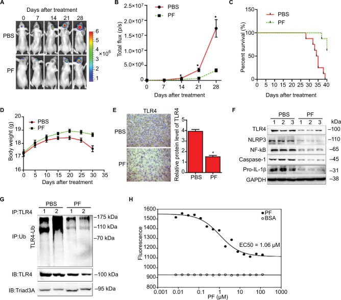
Figure 5.
Effects of paeoniflorin on the orthotopic xenograft mouse model and specific binding of paeoniflorin with TLR4 protein.
Notes: U87-luciferace cells (5×10⁵) were intracranially injected into the mid-right striatum of 8-week-old female BALB/c nude mice. Ten days postinjection, tumor formation was detected by bioluminescence imaging, and the mice were separated into the following 2 groups: a group comprising mice that were intraperitoneally injected with PBS (control) and a group comprising mice that were intraperitoneally injected with paeoniflorin (400 mg/kg/day). Tumor sizes were measured once every 7 days. Bioluminescence imaging was used to measure tumor volumes. (A) A representative tumor volume in each group is shown at each time point, n=6. (B) Tumor volumes were examined at each time point in each group, n=6. (C) The survival rate of each group, n=6. (D) Body weight changes in each group, n=6. At the end of the experiment or after the mice had died, the brains were removed (E) and were stained for TLR4 (scale bar=20 μm), n=3. The images were analyzed by Image-Pro-Plus. At the end of the experiment or after the mice had died, the tumor tissues were excised, and the protein lysates, n=3 (F) were used to estimate TLR4 signaling, n=3. (G) The lysates were immunoprecipitated with anti-TLR4 antibody and immunoblotted with antibodies against TLR4 and Triad3A, n=2. (H) MST assay was used to measure the binding between paeoniflorin and TLR4 protein, n=3. EC50 values were automatically calculated by the curve fitting. All tests were performed in triplicate. *P<0.05 compared with control.
Abbreviations: MST, microscale thermophoresis; TLR4, Toll-like receptor 4; IB, immunoblotting; PF, paeoniflorin; Ub, ubiquitin.

