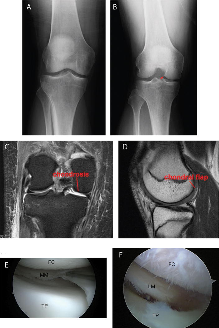
Fig. 1.

Examples of presence/absence of degenerative changes based on X-rays, MRI and arthroscopy. X-rays: (A) no OA; (B) joint space narrow and osteophyte formation (arrow) as evidence for early OA. MRI: (C) chondrosis; (D) chondral flap. Arthroscopy: (E) no chondrosis; (F) chondrosis. FC = femoral condyle, TP = tibia plateau, MM = medial meniscus, LM = lateral meniscus
