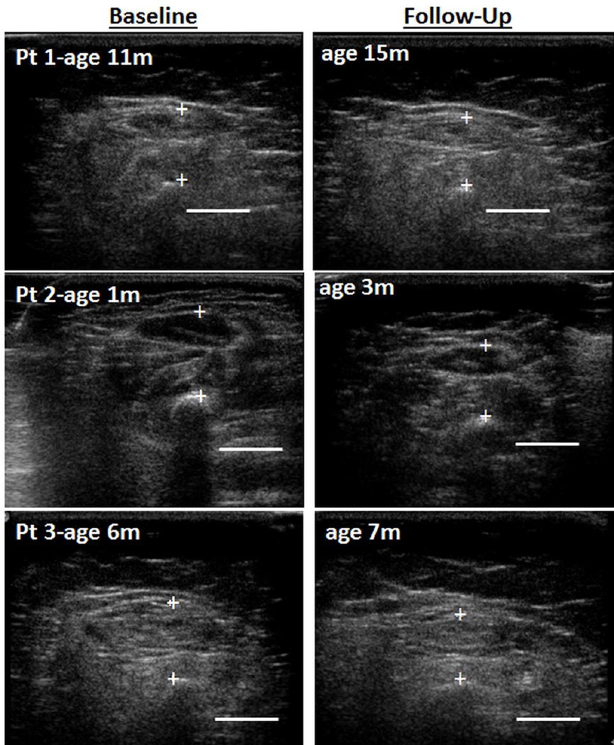
Figure 1. Ultrasound Images of Quadriceps in Three Children with SMA-1 Over Time.
Legend: Ultrasound cross sectional images of the quadriceps in three patients (Pt) with spinal muscular atrophy (SMA) type 1 shows muscle atrophy and worsening (increased) echointensity over time. Note the relatively normal muscle appearance of the youngest patient (#2, 1 month (m) old) at baseline (left panel) compared to the older patients. Bar = 1cm.

