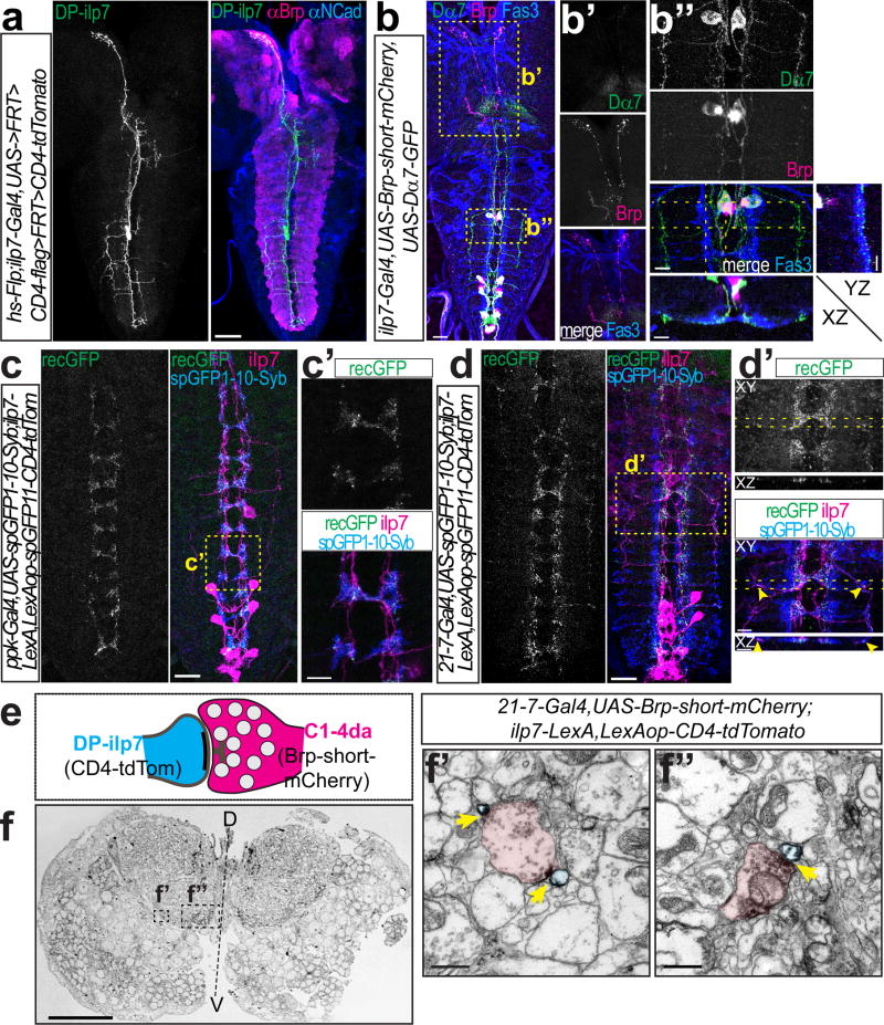
Figure 2. DP-ilp7 neurons are connected to multiple classes of sensory da neurons.
(a) DP-ilp7 neuron morphology visualized by Flp-mediated CD4-tdTomato labeling together with neuropil markers (anti-Brp in magenta, anti-N-Cadherin in blue). Scale bar: 50 µm. (b) Ilp7 neurons expressing presynaptic Brp-short-mCherry and postsynaptic Dα7-GFP markers together with anti-Fas3 labeling (Scale bar: 25 µm). Boxed regions show (b’) Brp-positive DP-ilp7 axon terminals in the PI region and (b”) Dα7-positive postsynaptic sites of DP-ilp7 neurons align with Fas3-labeled sensory terminals (YZ and XZ resliced projections indicated by dotted lines). Scale bars: 10 µm. (c) Syb-GRASP showing native reconstituted GFP signal (recGFP) between C4da (Syb-GFPsp1-10) and DP-ilp7 neurons (GFPsp11-CD4-tdTomato). Scale bar: 25 µm. (c’) Enlarged view of boxed region in (c). Scale bar: 5 µm. (d) Syb-GRASP between sensory da neuron expressed Syb-GFPsp1-10 (anti-GFPsp1-10) and DP-ilp7 neurons (GFPsp11-CD4-tdTomato) shows native reconstituted GFP (recGFP) along ventromedial and -lateral DP-ilp7 projections. Scale bar: 25 µm. (d’) Enlarged view of boxed region in (d). Arrowheads indicate lateral synapses of DP-ilp7 with C2da neurons. Scale bars: 5 µm. (e) Schematic of a sensory-DP-ilp7 neuron synapse visualized by DAB-labeled plasma membrane (CD4-tdTomato in ilp7) and sensory da neuron presynaptic active zone markers (Brp-short-mCherry). (f) Semi-thin VNC cross-section with specific DAB-labeling in the ventro-lateral and –medial neuropil. Scale bar: 25 µm. Ultrathin section of (f’) ventrolateral presynaptic terminals (C2da) contacting DP-ilp7 neurons and (f”) ventromedial contact between da neurons (C4da) and DP-ilp7 neurons. DAB-labeled active zones are indicated by arrows. Scale bars: 1 µm.

