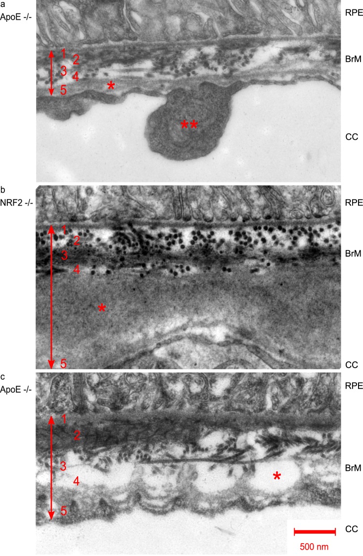
Figure 4.
TEM (12,000-fold magnification) of RPE/BrM/choroid complex pathologies in AMD mouse models. Numbers indicate BrM layers: 1, basal lamina of RPE; 2, inner collagenous layer; 3, elastic layer; 4, outer collagenous layer; 5, basal lamina of choriocapillaris. a single asterisk shows basal lamina splicing, double asterisk shows endothelial thickening, both typically found in AMD, b asterisk indicates outer collagenous layer deposits, and c asterisk indicates the formation of vacuoles within the outer collagenous layer.

