Figure 1.
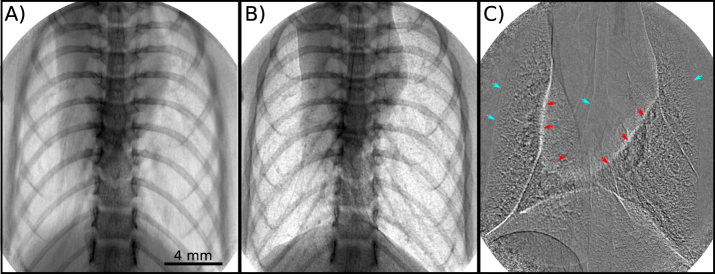
Propagation-based X-ray phase-contrast images of a mouse chest in vivo show the increase in image quality that is possible with sufficient flux to image the lungs without motion blur. In panel (A), an image was captured mimicking the exposure conditions at low-flux X-ray sources, resulting in motion blur due to the required averaging over breath cycles (here: one cycle during the 500 ms exposure). Panel (B) shows the benefit of a high flux source, which enables short exposures during plateaus of mechanical ventilation, in this case a 200 ms exposure triggered at the beginning of a 222 ms peak-inspiration breath-hold. Panel (C) reveals detailed structures in the lungs via a pseudo-differential image, achieved by subtracting sequential 200 ms breath-hold exposures between which there is a slight movement of the lungs and negligible movement of the skeleton. The red arrows highlight the border of the heart and the blue the edges of the lung and airways. The resulting structure seen within the lungs includes the small airways that are not easily seen in Panel (B).

