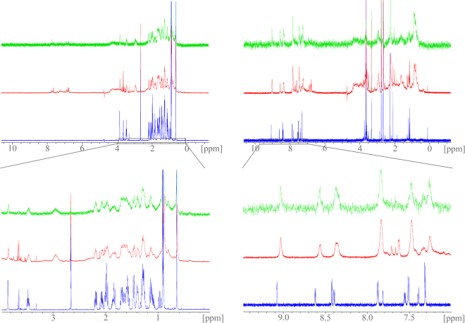
Figure 2.
Saturation-transfer difference (STD) NMR experiments confirm binding of 4 to the FXR-LBD: STD-NMR experiments with CDCA (1, left spectra) or imatinib (4, right spectra) and the FXR-LBD; the upper spectra are covering the full recorded range, the lower magnifications depict the relevant areas; standard 1H-NMR spectra of compound alone are depicted in blue, standard 1H-NMR spectra in presence of the FXR-LBD are red and STD-NMR spectra are green: 1H-NMR signals of 1 and 4 are significantly broadened upon addition of FXR-LBD protein. Additionally, signals of 4 show chemical shift perturbations. When the protein is saturated in the STD experiment (green), magnetization is transferred to 4 indicated by appearance of the same broad aromatic signals of 4 which confirms binding of 4 to the FXR-LBD.

