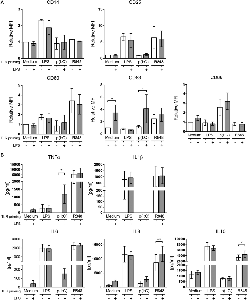
Fig. 2. TLR3 priming synergizes with low dose TLR4 for an increased inflammatory response by monocytes.
Monocytes were primed or not for 24 h with LPS, poly(I:C) or R848, then challenged with (blue bar) or without (white bar) with 0.2 ng/ml LPS for 5 h. a The surface expression of CD14, CD25, CD80, CD83 and CD86 were checked by flow cytometry (monocytes distinguished by FSC-A/SSC-A). The data are mean MFI ± SD relative to each control (non-stimulated monocytes) and accounts for more than three independent experiments. b TNFα, IL6, IL8, IL10 and IL1β levels found in culture supernatants of monocytes after LPS challenge. Data is mean ± SD and accounts for four independent experiments. Statistical differences towards LPS response are shown as *p < 0.05; **p < 0.01 by two-way ANOVA

