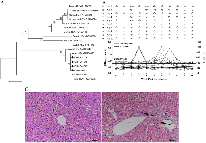
Fig. 1. Avian HEV gene analysis and clinical evaluation of the virus infected rabbits.
a Phylogenetic trees based on the sequences of four partial of HEV ORF2 genes (CHN-SN-C2, CHN-SN-D2, CHN-SN-G2, and CHN-SN-R2) isolated in this study and other HEVs from various animal species. The trees were generated by the neighbor-joining method with bootstrap tests of 1000 replicates using the MEGA 7.0 software. b Viremia/fecal viral shedding, ALT levels, and antibody levels in rabbits experimentally infected with CaHEV. c Microscopic lesions in livers from negative control rabbits (left) and rabbits experimentally infected with CaHEV (right) showed lymphocytic venous periphlebitis (arrows). Liver sections were stained with hematoxylin and eosin

