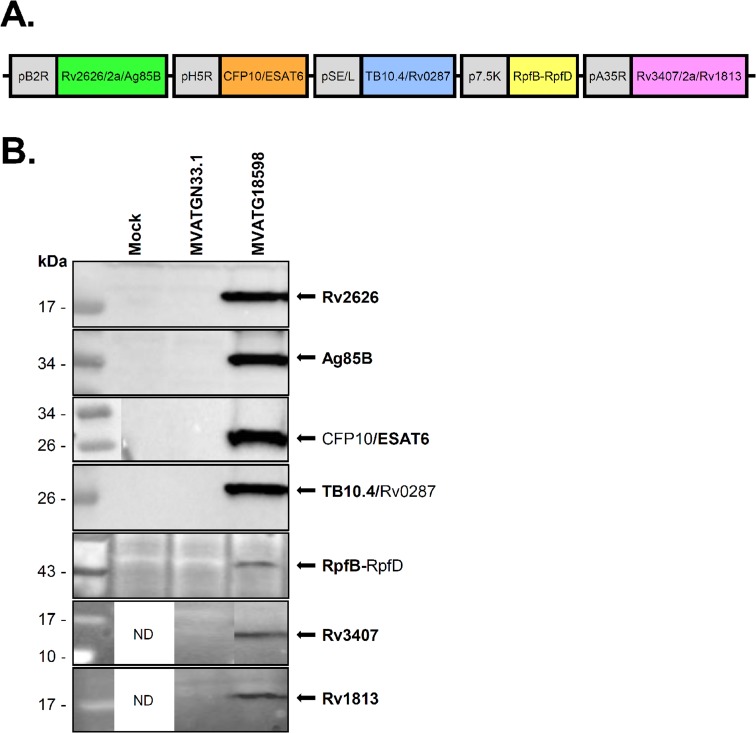
Fig 1. Schematic representation of antigenic expression cassettes in MVATG18598 and in vitro detection of antigen expression.
(A) MVATG18598 contains the fusion Rv2626/T2a/Ag85B under the control of pB2R promoter, the fusion CFP10/ESAT6 under the control of pH5R promoter, the fusion TB10.4/Rv0287 under the control of pSE/L promoter, the fusion RpfB-RpfD under the control of p7.5K promoter and the fusion Rv3407/E2a/Rv1813 under the control of pA35R promoter. (B) CEF cells were infected or not (Mock) with MVATG18598 or MVATGN33.1 and cell extracts analyzed by Western blot. Fusion Rv2626/T2a/Ag85B was detected using a mouse monoclonal anti-Rv2626 antibody (expected molecular weight of cleaved product: 17.4 kDa) and a rabbit polyclonal anti-Ag85B antibody (expected molecular weight of cleaved product: 31.3 kDa). CFP10/ESAT6 fusion (expected molecular weight: 21.8 kDa) was detected using a mouse monoclonal anti-ESAT6 antibody. TB10.4/Rv0287 fusion (expected molecular weight: 21.2 kDa) was detected using an anti-TB10.4 rabbit polyclonal antibody. Fusion RpfB-RpfD (expected molecular weight: 37.0 kDa) was detected using a rabbit polyclonal anti-RpfB antibody. Fusion Rv3407/E2a/Rv1813 was detected using a rabbit polyclonal anti-Rv3407 antibody (expected molecular weight of cleaved product: 13.2 kDa) and a rabbit polyclonal anti-Rv1813 antibody (expected molecular weight of cleaved product: 12.1 kDa). ND; not done.

