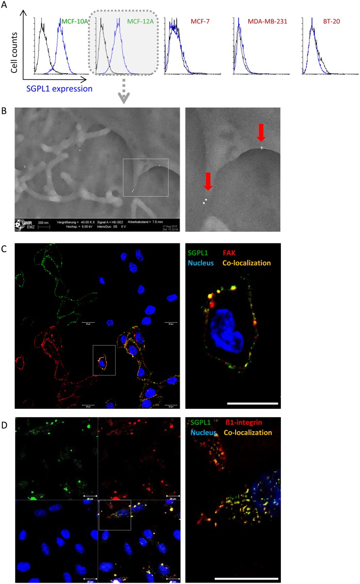
Fig 4. SGLP1 protein in association with the plasma membrane.
A: SGPL1 labeling on living breast cells quantitatively measured by flow cytometry. Only the non-tumorigenic breast cells MCF-10A and MCF-12A exhibit SGPL1 signals on the cell surface. B: Scanning electron microscopy of gold-labeled SGPL1 proteins on the cell surface of the non-tumorigenic breast cell line MCF-12A. C/D: Co-localization studies of SGPL1 and known plasma membrane adhesion molecules for MCF-12A C: focal adhesion kinase (FAK) and D: ß1-integrin. SGPL1: green, FAK or ß1-integrin: red, membrane protein overlay: yellow, nuclei: blue. Bar = 20 μm.

