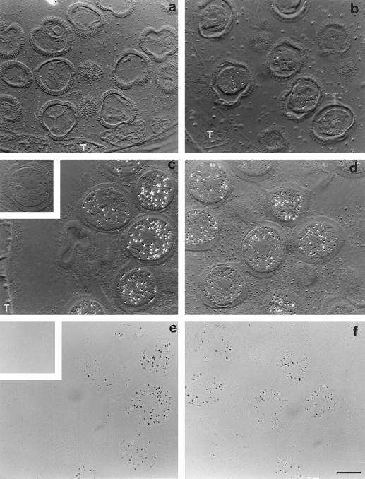
Figure 6.
Immunolocalization of BPC1 in the cytosol of developing pollen grains of B. napus W1. Sections of LR White embedded anthers were treated with mouse anti-rAPC1 followed by colloidal gold-tagged rabbit-anti mouse IgG and then silver enhanced. Silver grains, indicating the presence and location of BPC1, appear as bright refractive grains under differential interference contrast optics (a–d). a, Pollen grains from flower buds at the 4-mm stage; BPC1 protein was not detected at this stage. b, Pollen from flowers at the 6-mm stage; BPC1 protein is barely detectable, as illustrated by the sparse labeling in the four more prominent pollen grains in the micrograph. T, tapetum. c and d, Pollen from flowers at the 8-mm stage and the mature stage, respectively. There has been a marked increase in BPC1-labeling intensity. It is obvious that BPC1 is cytosolic, because neither pollen walls nor nuclei (c, grain center right; d, grains center and lower left) are labeled. e and f, Bright-field images of c and d, respectively, confirming the location of the label (silver grains appear as black grains) and demonstrating that the granular nature of the extracellular matrix seen in c and d is not due to labeling. The signal lies fully within the pollen grain walls and outside the nuclei. Inset in c and e show a grain from 8-mm stage flowers as a preimmune control. No labeling is evident in grains treated with preimmune mouse serum in place of anti-rAPC1. All micrographs are ×1000. Bar = 10 μm.

