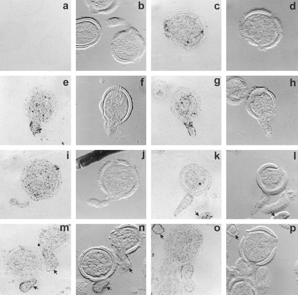
Figure 7.
Immunolocalization of BPC1 in germinated pollen grains of B. napus W1. Sections of embedded germinated grains were treated with mouse anti-rAPC, followed by colloidal gold-tagged rabbit-anti-mouse IgG, and were then silver enhanced. Bright-field and corresponding differential interference contrast images are presented in each case for clarity. a and b, Preimmune control images. c to p, Immune images. Note that signals are not restricted to the cytosol, but some labeling is also seen in the pollen grain wall. Labeling is intense on or near the surface of the pollen tubes, as evidenced by intense edge labeling, seen in e to h and k to p. Lack of intensity of edge labeling of the tube in I and j is probably due to the grazing nature of the section. Arrows in k, l, o, and p indicate pollen tubes of grains lying above or below the plane of section. Arrows in m to p further indicate the edge labeling. All images are ×1000.

