Fig. 5.
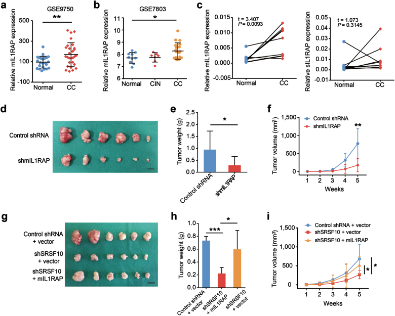
MIL1RAP participates in SRSF10-mediated CC tumorigenesis. a Analysis of mIL1RAP mRNA levels in normal cervical tissues and CCs in GSE9750 indicated in Fig. 2a. b Analysis of mIL1RAP mRNA levels in normal cervical tissues, CINs, and CCs in GSE7803 indicated in Fig. 2c. c MIL1RAP expression was examined by RT-PCR in 10 pairs of human CC and adjacent non-cancerous tissues. sIL1RAP was detected as a control. d Ms751 cells were infected with either retrovirus expressing mIL1RAP-knockdown shRNA (shmIL1RAP) or control shRNA and selected for puromycin resistance. Stable Ms751 cells were injected into nude mice which were randomly divided into two groups (n = 6). Mice were killed at 5 weeks after injection. Tumors were excised from the mice and weighed (scale bar: 1 cm). e Results are shown as mean ± s.d. of the tumor weight. f Time-course of xenograft growth. Tumor volumes of the mice described in d were measured every week. Each point represents the mean ± s.d. of the tumors. g Ms751/shSRSF10 or shRNA control cells were transfected with either retrovirus expressing mIL1RAP or empty-vector control (vector). Stable Ms751 cells were injected into nude mice which were randomly divided into three groups (n = 6, scale bar: 1 cm). h Results are shown as mean ± s.d. of the tumor weight. i Time-course of xenograft growth. The tumor volumes of mice described in g were measured every week. Each point represents the mean ± s.d. of the tumors. *P < 0.05; **P < 0.01; ***P < 0.001

