Fig. 3.
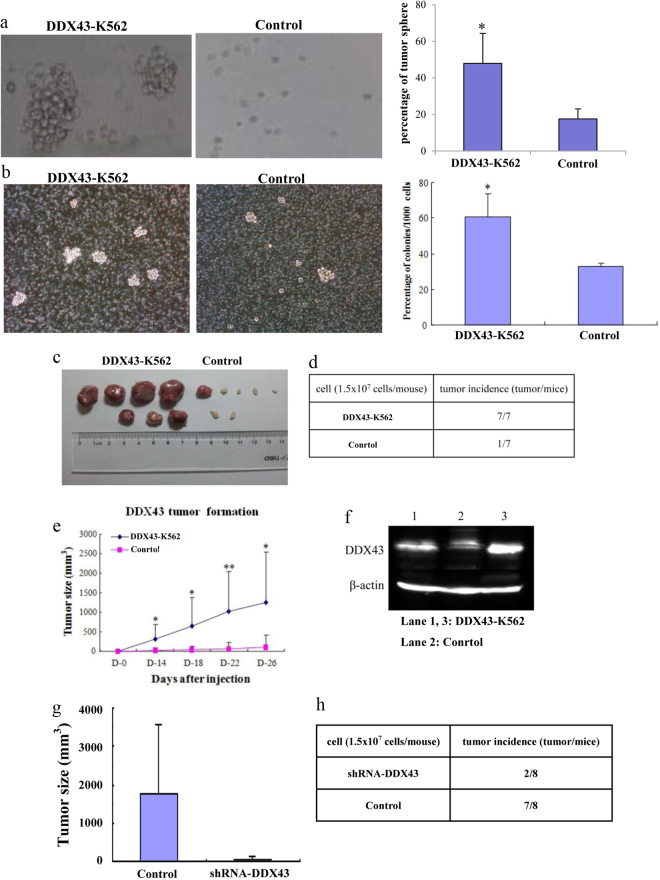
DDX43 overexpression affected clone formation and tumor formation. a Firstly, 2% agarose was plated into six-well plates, and DDX43-K562 or control cells were mixed with 0.3% low melting agarose, respectively. More and larger colonies were observed in the DDX43-K562 group than in the control group after 4 weeks. b K562 cells stably transfected with DDX43 or the control vector were plated in 1% methylcellulose with 10% FBS. c, d DDX43- or mock-transfected K562 cells were inoculated into CD1 strain nude mice subcutaneously. Mice were photographed and killed after 4 weeks of injection. e We measured tumor sizes and the tumor growth curves were drawn. Data showed the mean ± SD from three experiments. f DDX43 protein detected by western blot in tumors from mice inoculated with DDX43-K562 or control cells. g K562 cells transfected with DDX43 shRNA-1 or a control vector were inoculated into CD1 strain nude mice subcutaneously. Mice were photographed and killed after 4 weeks of injection. Tumor sizes were measured. h Tumor incidence from K562 cells transfected with DDX43 shRNA-1 or a control vector. *P < 0.05; **P < 0.01

