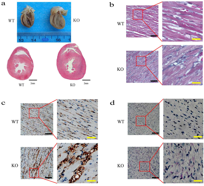
Figure 2.
Histopathology of myocardium from Lamp2y/− rats and wild-type rats (8 weeks). (a) Gross morphology of the heart; (b) Masson Trichrome staining; (c) Immunohistochemical reactivity for MAb B7, a marker for myocardium microvascular endothelial cells32; (d) Periodic Acid Schiff (PAS) staining. Bars in (a) correspond to 2 mm. The black bars correspond to 200 μm, and the yellow bars correspond to 2 mm in (b,c and d). WT, wild-type male rats; KO, Lamp2y/− rats.

