Figure 5.
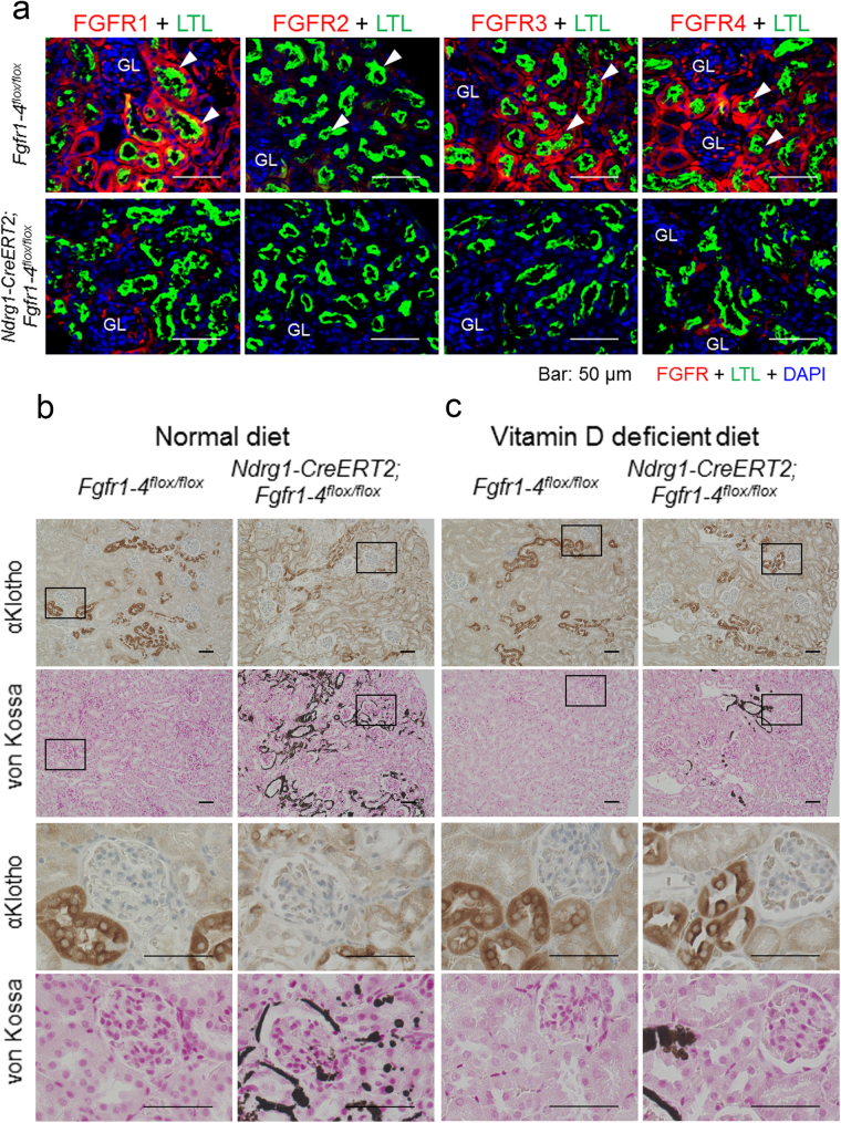
Proximal tubule-specific deletion of FGFR1–4 induces ectopic calcification and loss of αKlotho expression in distal tubules, both of which can be reversed by a vitamin D-deficient diet. (a) Indirect immunofluorescence staining studies for FGFR1, FGFR2, FGFR3, and FGFR4 in Fgfr1–4flox/flox and Ndrg1-CreERT2;Fgfr1–4flox/flox mice at 3 weeks after tamoxifen treatment. Kidney sections were immunostained for FGFR (red) with marking of proximal tubules with LTL (green) and counterstaining of cell nuclei with DAPI (blue). GL: glomerulus. Scale bars: 50 μm. (b) Proximal tubular ablation of FGFRs in mice maintained on a normal diet causes cortical ectopic calcification, disassembly of distal tubular cells, and loss of distal tubular αKlotho expression. Kidney serial sections were stained for αKlotho and calcium deposition using DAB-based immunostaining and von Kossa staining methods, respectively. Rectangular areas are magnified below in the two rows (αKlotho and von Kossa). Scale bars: 50 μm. (c) Reversal of cortical calcification, disassembly of distal tubular cells, and loss of distal tubular αKlotho expression in mice on a vitamin D-deficient diet. The experiments were carried out and shown as in (b). Kidney cortical surface is oriented to the right. The same experiments were repeated in 3 different littermates with and without the Ndrg1-CreERT2 transgene under the C57BL/6 J Fgfr1–4flox/flox background. Shown are representative micrographs. See also Supplementary Figure S3.

