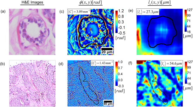
Figure 2.
Computing the geometric feature and scattering feature over each annotated gland. (a) and (b) H&E images of benign and malignant glands, respectively. (c) and (d) SLIM images of the same benign and malignant glands, respectively, illustrating gland curvature . The median over gland is used as the geometric feature for classification. (e) and (f) for benign and malignant glands, respectively. The median over gland is used as the scattering feature for classification.

