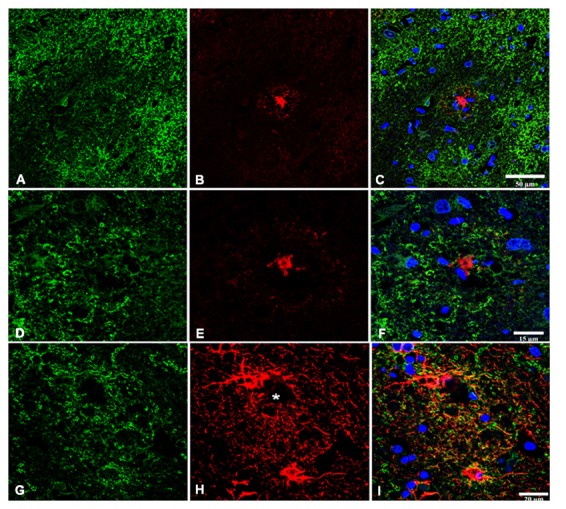
Figure 3.

Double-labeling immunofluorescence and confocal microscopy of GLT1 (green), β-amyloid (red; B,C,E,F), and GFAP (red; H,I) in frontal cortex area 8 in AD. GLT1 immunoreactivity is preserved in the vicinity of β-amyloid plaques (A–F). Astrocytes surrounding small β-amyloid deposit (asterisk) show preserved GLT1 immunoreactivity at the cell membrane (G–I). Paraffin sections, nuclei (blue) are stained with DRAQ5™; (A–C), bar = 50 μm; (D–F), bar = 15 μm; (G–I), bar = 20 μm.
