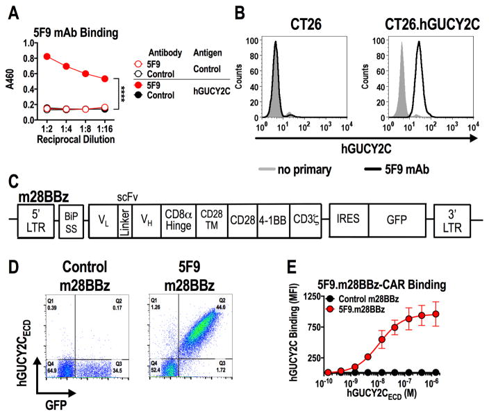
Figure 1. Generation of human GUCY2C-specific CAR-T cells.
(A) Recombinant 5F9 antibody was assessed by ELISA for specific binding to hGUCY2CECD or BSA (negative control) plated at 1 μg/mL. Two-way ANOVA; ****p<0.0001. (B) Flow cytometry analysis was performed on parental CT26 mouse colorectal cancer cells or CT26 cells engineered to express hGUCY2C (CT26.hGUCY2C) and stained with 5F9 antibody. (C) Schematic of the third-generation murine CAR construct containing murine sequences of the BiP signal sequence, 5F9 scFv, CD8α hinge region, the transmembrane and intracellular domain of CD28, the intracellular domain of 4-1BB (CD137), and the intracellular domain of CD3ζ (5F9.m28BBz). The CAR construct was inserted into the MSCV retroviral plasmid pMIG upstream of an IRES-GFP marker. (D) Murine CD8+ T cells transduced with a retrovirus containing a control (1D3.m28BBz) CAR or CAR derived from the 5F9 antibody (5F9.m28BBz) were labeled with purified 6xHis-hGUCY2CECD (10 μg/mL), detected with anti-5xHis-Alexa Fluor 647 conjugate. Flow plots were gated on live CD8+ cells. (E) 6xHis-hGUCY2CECD binding curves for 5F9-derived or control (1D3) CARs, gated on live CD8+GFP+ cells (Supplementary Fig. S5). Combined from 3 independent experiments.

