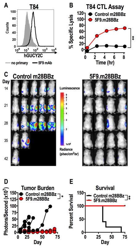
Figure 4. hGUCY2C CAR-T cells eliminate human colorectal tumor xenografts.
(A) hGUCY2C expression on T84 human colorectal cancer cells was quantified by flow cytometry using the recombinant 5F9 antibody. (B–E) Control (1D3.m28BBz) or 5F9.m28BBz CAR constructs were transduced into murine CD8+ T cells. (B) T84 colorectal cancer cells in an E-Plate were treated in duplicate with 5F9-m28BBz or control CAR-T cells (5:1 E:T ratio), media, or 10% Triton-X 100 (Triton), and the relative electrical impedance was measured every 15 minutes for 20 hours to quantify cancer cell death (normalized to time=0). Percent specific lysis values were calculated using impedance values following the addition of media and Triton for normalization (0% and 100% specific lysis, respectively). Two-way ANOVA; **p<0.01; representative of two independent experiments. (C–E) Immunodeficient NSG mice were injected with 2.5x10⁶ luciferase-expressing T84 colorectal cancer cells via intraperitoneal injection and were treated with 107 control (N=5/group) or 5F9-m28BBz (N=4/group) CAR-T cells on day 14 by intraperitoneal injection. (C–D) Total tumor luminescence (photons/second) was quantified just prior to T-cell injection and weekly thereafter. Two-way ANOVA; *p<0.05. (E) Mice were followed for survival. Log-rank Mantel Cox test; *p<0.05.

