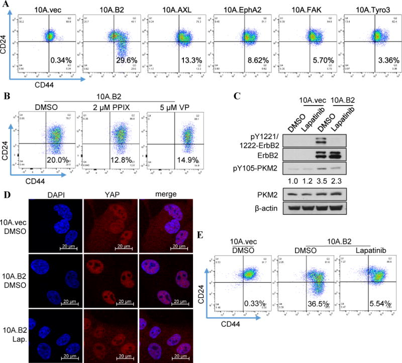
Figure 5. Abrogation of YAP downstream signaling inhibited oncogenic kinase-induced cancer stem-like cell population in vitro. A).

Flow cytometry analysis of the cancer stem-like cell populations (CD44hi/CD24neg) in MCF10A cells transfected with expression vectors of indicated kinases that can phosphorylate PKM2-Y105. B) Flow cytometry analysis of the cancer stem-like cell population (CD44hi/CD24neg) in the 10A.B2 cells treated with DMSO (as controls), 2 μM PPIX, or 5 μM Verteporfin. C) Western blot analysis of the pY1221/1222- ErbB2, total ErbB2 protein, pY105-PKM2, and total PKM2 protein levels in the 10A.vec and 10A.B2 cells after treatment with DMSO or lapatinib (1 μM) for 24h. Quantification of pY105-PKM2 comparing different samples with 10A.vec.DMSO (normalized by β-actin) was conducted by Image J software. D) Representative immunofluorescent staining showing the colocalization of YAP protein (red) and nuclear (DAPI, blue) in the DMSO-treated 10A.vec cells and the DMSO- or lapatinib-treated (1 μM) 10A.B2 cells. E) Flow cytometry analysis of the cancer stem-like cell population (CD44hi/CD24neg) in the DMSO-treated 10A.vec cells and the DMSO- or lapatinib-treated (1 μM) 10A.B2 cells.
