
Figure 2. PARP-1 siRNA reduces cell proliferation and induces apoptosis in HER2+ trastuzumab resistant breast cancer cell lines.
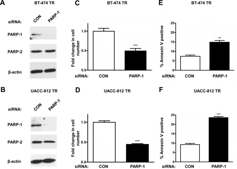
BT-474 TR and UACC-812 TR were transfected with 20 nM of control (CON) or PARP-1 siRNA for 96 hours. Knockdown of PARP-1 protein expression levels was verified via Western Blot analysis. β-actin was used as a loading control (A and B). Data shown are representative immmunoblots from one of three independent experiments. Following PARP-1 knockdown, cell counts were obtained via cellular proliferation assays (C and D). Apoptosis was assessed with FACS analysis using propidium iodide and Annexin V staining (E and F). The representative figures shown are from one of three independent experiments performed in (C and D) quadruplicate or (E and F) triplicate. ***p≤0.0005, ** p<0.005