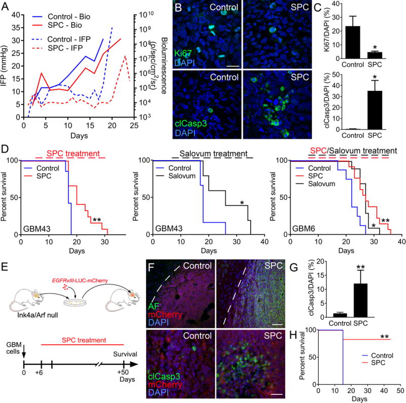
Fig. 3. AF treatment reduces tumor growth in GBM models.

(A) Parallel IFP and bioluminescence measurements showed that treatment with SPC diet reduced IFP levels and tumor growth compared to control GBM43 xenografts. (B–C) SPC treatment reduced proliferation (Ki67) and induced apoptosis (cleaved caspase 3) in GBM43 xenografts. Scale bar: 30 μm. (n=3, *P < 0.05, Student’s t test) (D) Kaplan-Meier curves showing increased survival in GBM43 xenografts treated with SPC diet (n=7 for control and n=12 for SPC, **P < 0.01; Log-rank Mantel-Cox test) or Salovum (n=7 for control and n=5 for Salovum, *P < 0.05; Log-rank Mantel-Cox test) compared to control. SPC diet and Salovum also increased survival in EGFRvIII-expressing GBM6 xenografts (n=8 for control, n=13 for SPC, and n=10 for Salovum *P < 0.05, **P < 0.01; one-way ANOVA). (E) Immunocompetent GBM model based on established neurospheres from the subventricular zone of Ink4a/Arf null FVBn mice that were lentivirally transduced to express human EGFRvIII, mCherry, and LUC. Mouse GBM cells were allografted into adult FVBn mice and administered SPC diet on day 6. (F–G) SPC treatment resulted in AF induction in tumor cells (dotted line; tumor border) and induction of apoptosis (cleaved caspase 3). Scale bar: top panel 50 μm, bottom panel 20 μm. (n=3, **P < 0.01, Student’s t test). Kaplan-Meier (n=5 for control and n=5 for SPC, **P < 0.01; Log-rank Mantel-Cox test) curve showing increased survival following SPC treatment in GBM43 xenografts.
