Figure 4.
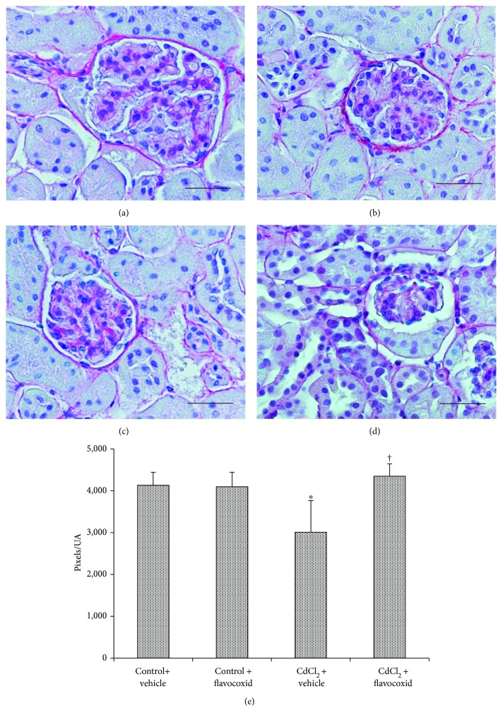
Structural organization of the interstitial connective tissue of kidneys from mice of control plus vehicle (0.9% NaCl, 1 ml/kg/day i.p.), control plus flavocoxid (20 mg/kg/day i.p.), CdCl2 (2 mg/kg/day i.p.) plus vehicle, and CdCl2 plus flavocoxid groups (Sirius red stain). (a, b) In both control plus vehicle and control plus flavocoxid-treated mice, the normal presence of collagen fibers is evident in the interstitial tissue. (c) In CdCl2-challenged mice, SR stain is less evident around the glomerular capsule and the tubules. (d) In CdCl2 plus flavocoxid-treated mice, no apparent difference with normal specimens is present. (e) Quantitative evaluation of the SR-positive areas. ∗p < 0.05 versus both controls and †p < 0.05 versus CdCl2 plus vehicle (scale bar: 50 μm).

