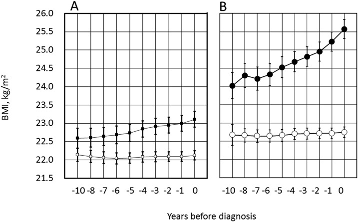
Figure 3.
Trajectories of BMI before diagnosis of (A) PDM and (B) diabetes. Symbols are the same as in Fig. 2. (A and B) Values in the progressors and nonprogressors at each time point were significantly different both in A and B (P < 0.01 for each). The axis scale was intentionally maintained the same to facilitate visual comparison. See Fig. 2A and 2B for the number of individuals examined each year.

