Figure 3.
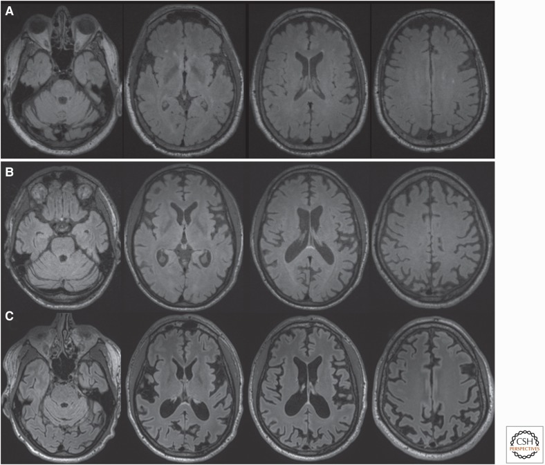
Brain magnetic resonance images (MRIs) in Slow forms of genetic prion disease (gPrD). (A) Fluid-attenuated inversion recovery (FLAIR) brain MRI in a 57-yr-old patient with P102L gPrD possibly 16.5 yr after onset (precise age of onset unclear because of comorbidities) shows mild cerebellar atrophy but no cortical atrophy or typical T2-weighted or diffusion weighted imaging (DWI) hyperintensites (cortical ribboning or deep nuclei hyperintensity), but only several punctate hyperintensities in the white matter consistent with small vessel ischemic vascular disease. DWI/attenuation diffusion coefficient (ADC) map sequences showed no restricted diffusion (not shown). (B) FLAIR brain MRI of with 37-yr-old gPrD 6-octapeptide repeat insertion (OPRI) patient showing diffuse cortical atrophy only 7 mo after reported clinical onset; the amount of atrophy suggests that the disease began years before obvious clinical onset. DWI and ADC map sequences showed no restricted diffusion (not shown). (C) Repeat brain MRI on the same 6-OPRI patient 25 mo later, 32 mo after clinical onset, shows the progression of atrophy. DWI and ADC map sequences still showed no restricted diffusion (not shown). Orientation is radiological. (Reprinted from Takada et al. 2017.)

