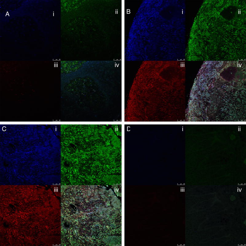
Figure 2.

Immunostaining of human gingival connective tissue for carbamylated, citrullinated and MAA-modified proteins. 2A; healthy gingival tissue, 2B; mild periodontitis, 2C; moderate periodontitis. 2D Isotype control showing no background signal. (i) = Carbamylated proteins (blue staining), (ii) = MAA-modified proteins (green staining, bottom left); (iii) = citrullinated proteins (red staining), and (iv) = merged images of staining for all three modified proteins. Magnification: 40×; Bar = 50μm.
These sections correspond to the connective tissue fields shown in Figure 1.
