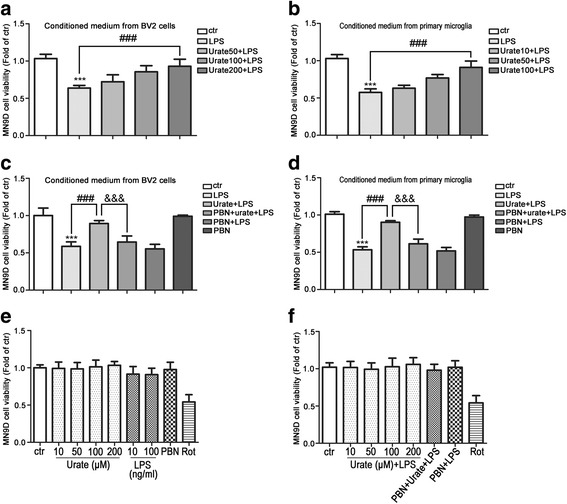
Fig. 5.

Urate protects DA neurons from neurotoxicity induced by microglia activation. a, b MN9D cells were incubated for 24 h with conditioned medium from cultures of BV2 cells (a, n = 4) or rat primary microglia (b, n = 4) treated with urate plus LPS. Cell viability was measured with the MTS assay. c, d MN9D cells were incubated for 24 h with conditioned medium derived from cultures of BV2 cells (c, n = 4) or rat primary microglia (d, n = 4) treated with LPS, urate+LPS, or PBN+urate+LPS. Cell viability was evaluated with the MTS assay. e, f MN9D cells were treated with indicated drugs for 24 h, and cell viability was evaluated with the MTS assay (n = 3). Rotenone (Rot, 0.5 μM) was used as positive control. Medium from cultures of untreated microglia served as a control (ctr). Data represent the mean ± SD. ***p < 0.001 vs. control group; ###p < 0.001 vs. LPS group; &&&p < 0.001 vs. urate+LPS group (one-way analysis of variance)
