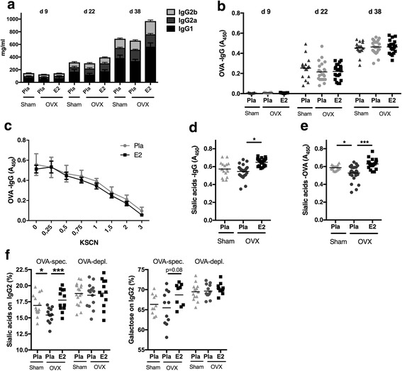
Fig. 1.

Estrogen influences immunoglobulin G (IgG) sialylation. Mice were ovariectomized (OVX) at 3 months of age, followed by insertion of slow-release treatment pellets with placebo (Pla) or estrogen (E2; 0.83 μg/day). Ten days after ovariectomy, the animals were immunized with ovalbumin (OVA), and 14 days later, they were boostered. Serum was taken at day 9 (d9) before immunization, day 22 (d22) after immunization, and day 38 (d38) at termination after immunization and boostering. a Total IgG concentrations: significant induction in all treatment groups after immunization (day 9–day 22) and after boostering (day 22–day 38) for all IgG subtypes. Data are mean ± SEM (n = 11–13). b Concentration of ovalbumin-specific IgG (OVA-IgG) (n = 17–24). c Titers of ovalbumin-specific IgG measured at termination (n = 10–12). d Concentration of sialic acids on total IgG measured at termination (n = 17–24). e Concentration of sialic acids on OVA-specific IgG measured at termination (n = 17–24). f Mass spectrometry-based analysis of sialic acids and galactose on IgG-Fc of OVA-specific (OVA-spec) and OVA-depleted (OVA-dep) IgG2 measured at termination (n = 12–14). Values are indicated on a scatter dot plot with mean indicated by a bar. Statistical analysis with analysis of variance followed by the Bonferroni multiple-comparisons test for selected time points. *p < 0.05, ***p < 0.001
