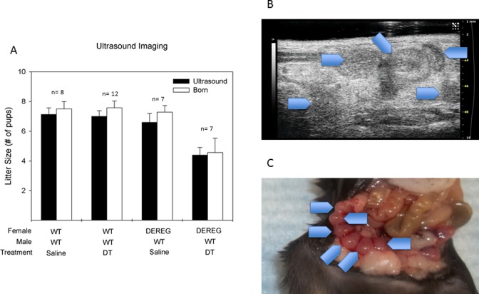
Figure 5.
Equivalent ultrasound visualized implantations to delivered pups demonstrated no postimplantation defects. (A) Comparison of ultrasound-visualized implanted pups and delivered pups. (B) Representative ultrasound picture of implanted embryos. Gestational age e+7. Blue arrowheads depict ultrasound-visualized implantations. (C) Representative picture of implanted embryos from same mouse depicted in Figure 4B. Blue arrowheads depict gestational sacs in the right uterine horn. Paired t test was used for analysis.

