Figure 2.
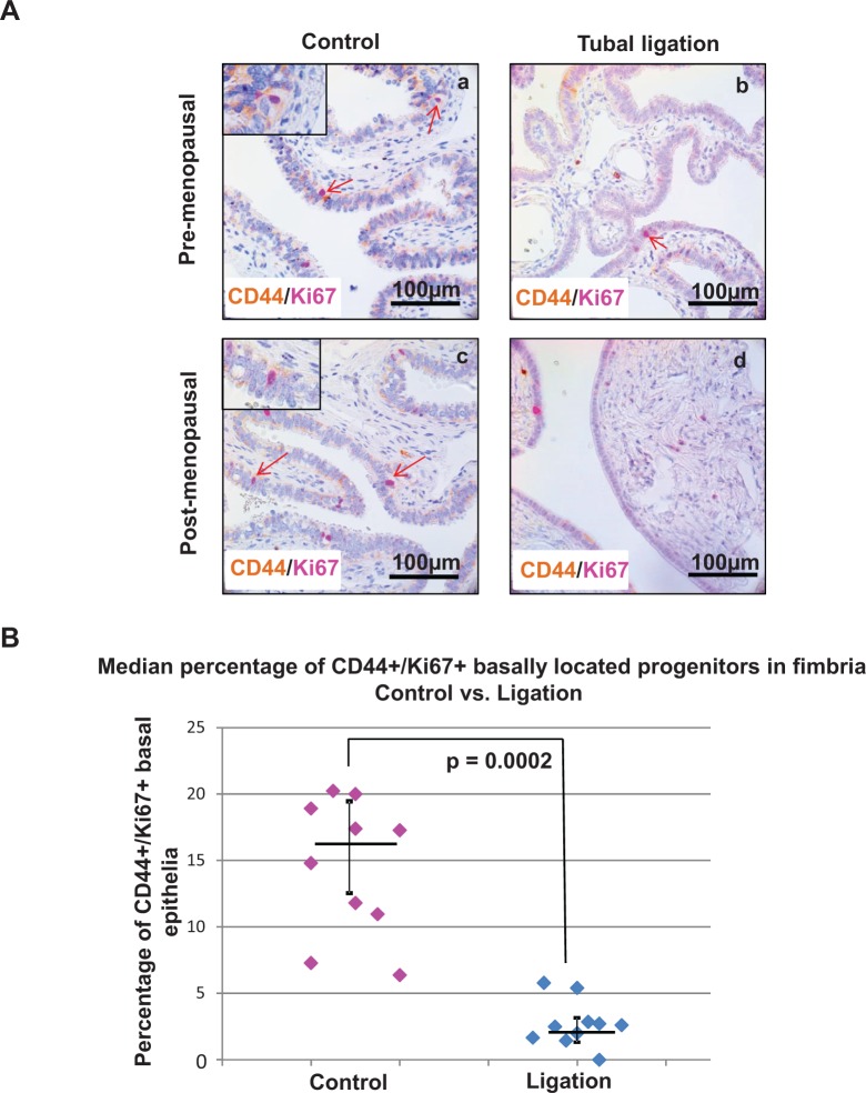
The percentage of proliferating epithelial progenitors was diminished in the distal fallopian tube of patient samples with tubal ligation. (A) Immunohistochemistry revealed a lower expression of proliferating progenitors (basally located CD44/Ki67 dual-positive epithelial cells) in the distal fallopian tube of ligated patient samples (b and d) compared to intact fallopian tubes (a and c). Light brown staining corresponds to CD44 expression and magenta staining indicates Ki67 nuclear expression. Tubal ligation samples from both pre- (b) or postmenopausal (d) patients showed a reduction in CD44/Ki67 dual expression compared to the control cohort. (B) The median percentage of proliferating progenitors was significantly lowered in the fimbria of tubal ligated patient specimens compared to age-matched controls at P = .0002. Each dot on the chart represents the percentage of basally located CD44-positive epithelial cells that also expressed Ki67. Horizontal bars represent the median for each cohort and the vertical bars denote interquartile range.

