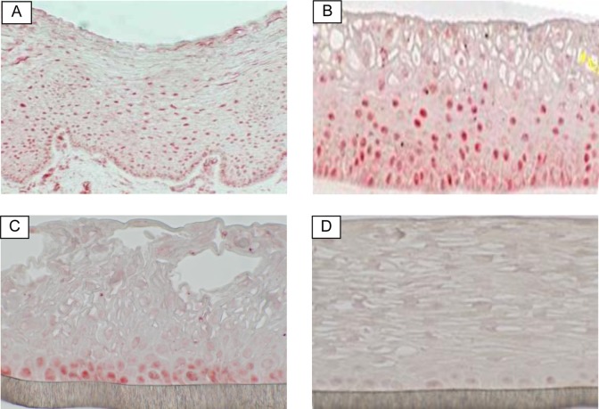
Figure 1.
Immunohistological staining of estradiol receptor (red) in: (A) human vaginal-ectocervical tissue explant (positive control), (B) full-thickness vaginal-ectocervical (VEC-FT) tissue model, (C) partial thickness vaginal-ectocervical tissue model (VEC-PT), and (D) VEC-PT tissue negative control (without primary antibody). (The color version of this figure is available in the online version at http://rs.sagepub.com/.)

