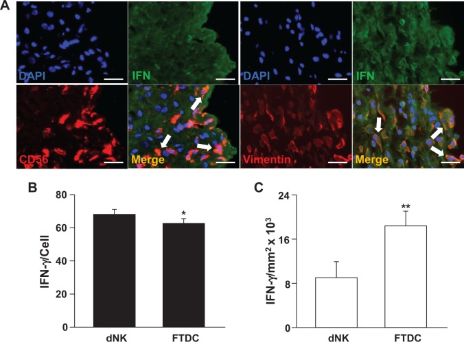
Figure 1.
Immunoreactive IFN-γ in decidual natural killer (dNK) cells and first-trimester decidual cells (FTDCs) in human decidua. A, First-trimester decidual sections were stained with either antihuman CD56 (red) or antihuman vimentin (red) antibodies and then incubated with rabbit antihuman IFN-γ (green). Staining with antihuman 4′,6′-diamidino-2-phenylindole (DAPI; blue) denotes nuclei. Arrows in merged images indicate yellow-orange immunofluorescence resulting from double staining. B, Ordinate indicates immunoreactive IFN-γ levels/CD56+ cell. C, Ordinate indicates IFN-γ levels/decidual mm2 × 103. The results are reported as mean ± standard error of the mean (SEM). n = 18; *P < .05 and **P < .01. Magnification: 400×; scale bar: 50 μm. IFN-γ indicates interferon γ.

