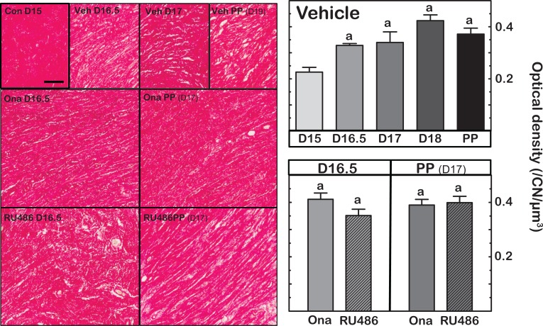
Figure 1.
Left panels are representative photomicrographs of Picrosirius red-stained collagen in sections of cervix obtained on specified days postbreeding. PP indicates postpartum. Group designations are intact controls (Con), vehicle-injected (Veh), and mice given PR antagonist, onapristone (Ona), or mifepristone (RU486) on day 16.5 postbreeding, respectively, and prepartum 8 hours after treatment. Right panels are graphs of optical density (OD; mean ± standard error of the mean [SEM]; n = 3-10) of polarized light from birefringence of Picrosirius red–stained sections. Data were normalized to cell nuclei density/section to account for variability in the area of extracellular space, cell size, cell numbers, and morphology across sections, individuals, and groups. The term collagen degradation reflects disarray in collagen cross-linked fibers and possibly content/area as described in the Methods section. a P < .05 versus D15 Vehicle (analysis of variance [ANOVA] with Dunnett test).

