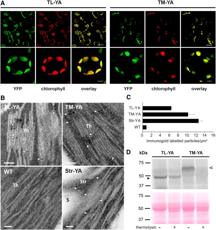
Figure 3.
Confocal microscopy analyses, immunogold labeling, and biochemical analyses of Arabidopsis seedlings of the TL-YA and TM-YA lines. A, Fluorescence microscopy images of mesophyll cells of Arabidopsis seedling leaves with a YFP filter and a chlorophyll filter. An overlay of the two channels also is shown. Bars = 25 µm (top row) and 5 µm (bottom row). B, Immunocytochemical analyses of aequorin subcellular localization. The wild-type (WT) line and a line expressing YA in the stroma (Str-YA) were used as negative and positive controls, respectively. White arrowheads indicate gold particles. Bars = 100 nm. S, Starch granule; Str, stroma; Th, thylakoids. C, Quantitative analyses of immunogold-labeled particles. Data are means ± se of 40 different fields from three biological replicates. D, Immunoblot analyses of isolated thylakoids, incubated in the absence (−) or presence (+) of 0.1 µg µL−1 thermolysin, as indicated. Isolated thylakoids corresponding to 50 µg of protein were probed with an anti-aequorin antibody (top gel). Equal loading was confirmed by Ponceau Red staining of the blot membrane (bottom gel). Black and white arrowheads indicate YA chimeras targeted to the thylakoid lumen and the thylakoid membrane, respectively.

