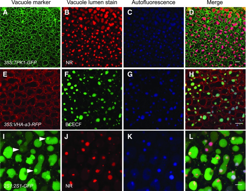
Figure 5.
Forming PSV are identified by the acidotropic stains NR and BCECF-AM and PSV luminal autofluorescence in bent cotyledon embryos. A to H, NR and BCECF-AM stain vacuoles labeled with the tonoplast markers TPK1-GFP (A) and VHA-a3-RFP (E), respectively. Vacuole lumen autofluorescence (blue) colocalizes with the stains (D and H). I to L, Embryos accumulating 2S1 albumin-GFP were stained with NR. The 2S1-GFP signal fills vacuole lumina, and areas of more intense GFP fluorescence are observed (arrowheads in I). NR stains distinct subregions of the vacuole lumina (J). These NR-stained subregions colocalize with PSV lumen autofluorescence (K) and with areas of intense 2S1-GFP fluorescence (L). Bars = 10 μm (A–H) and 5 μm (I–l).

