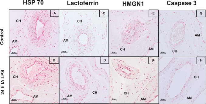
Figure 2.
Expression of several DAMPs in fetal membranes did not increase after IA LPS exposure: representative photomicrographs (20× objective) of the immunohistochemistry for HSP 70 showed staining in the amnion (AM) and chorion (CH) in control (A) and 24 hours after IA LPS exposure (B); lactoferrin in control (C) and 24 hours after IA LPS exposure (D); high mobility group nucleosome binding domain 1 (HMGN1) in control (E) and 24 hours after IA LPS exposure (F); and caspase 3 in control (G) and 24 hours after IA LPS exposure (H). No differences were observed in the expression of these DAMPs. DAMP indicates damage-associated molecular pattern; IA, intra-amniotic; LPS, lipopolysaccharide.

