Figure 1.

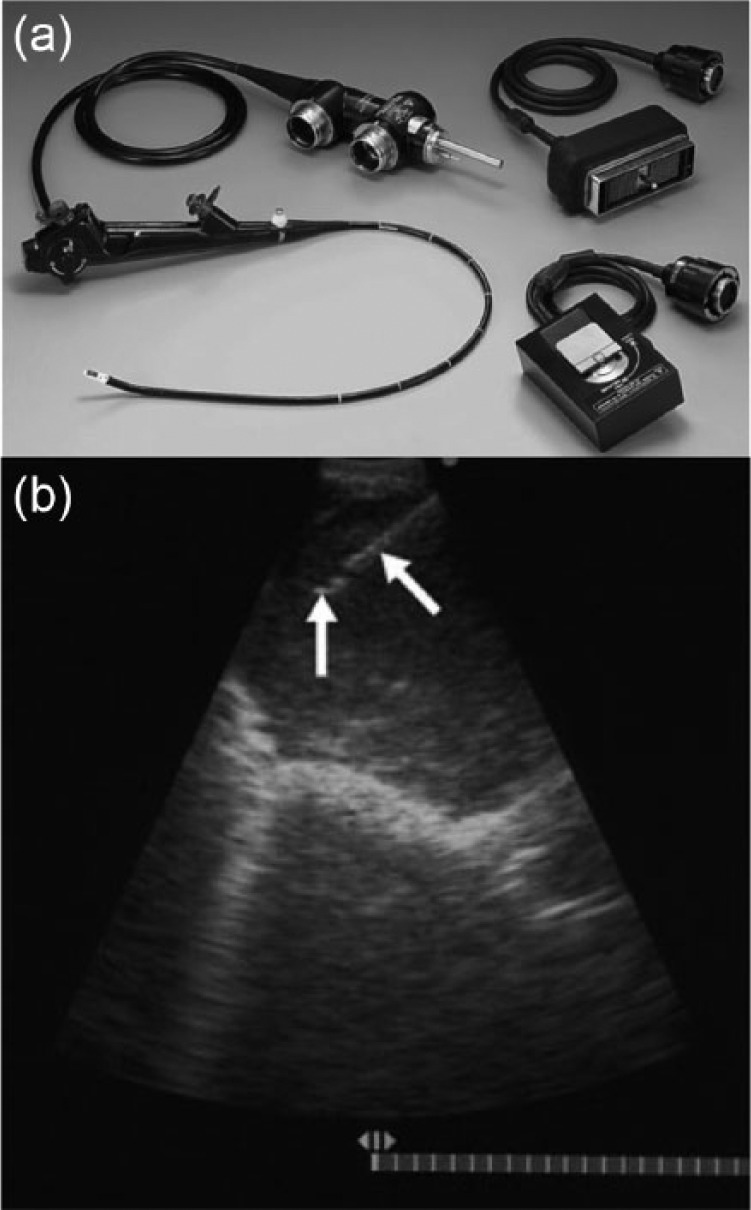
Convex endobronchial ultrasound (EBUS). (a) EBUS scope with associated white light and ultrasound cables. (b) Intraprocedural view of a biopsy. The white arrows point to the needle. Images © Georg Thieme Verlag KG.

Convex endobronchial ultrasound (EBUS). (a) EBUS scope with associated white light and ultrasound cables. (b) Intraprocedural view of a biopsy. The white arrows point to the needle. Images © Georg Thieme Verlag KG.