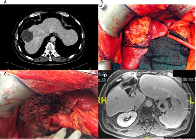
Figure 1.
Anatomic central hepatectomy (mesohepatectomy) for HCC. A, Venous phase of abdominal CT showing the centrally located tumor with involvement of the middle and right hepatic veins. The patient had a right inferior hepatic vein, which allowed the plans for a central hepatectomy. B, Intraoperative image of the tumor centrally located in the context of cirrhotic liver. C, Intraoperative image after central hepatectomy showing the central defect and spared right posterior and left lateral sections. D, Delayed phase of abdominal MRI 1 year after resection revealing central defect and enlarged right posterior and left lateral sections. CT indicates computed tomography; HCC, hepatocellular carcinoma; MRI, magnetic resonance imaging.

