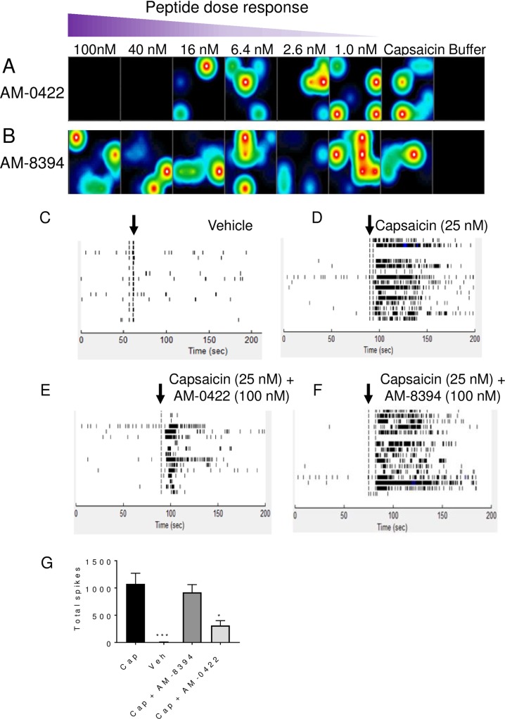
Fig 7. Inhibition of capsaicin-induced action potential firing in MEA recordings in rat DRG neurons by active peptide AM-0422 but not inactive peptide AM-8374.
Heat maps of active electrodes detecting action potential firing 30 seconds following vehicle, 25 nM capsaicin, 25 nM capsaicin plus 1–100 nM of AM-0422 (A) or 25 nM capsaicin plus 1–100 nM of AM-8394 (B). The colors white/red, yellow/green, and blue/black correspond to high, medium, and low action potential firing frequency respectively. Whole well raster plots for buffer (C), 25 nM capsaicin (D), 25 nM capsaicin plus 100 nM AM-0422 (E), or 25 nM capsaicin plus 100 nM AM-8394 (F) added at the time point indicated by the vertical black arrows. Each row in the raster plots represents a single electrode with 16 electrodes per well and all recordings were performed at 37°C. G. Summary of total spikes from active wells following 2 minute treatment with 25 nM capsaicin (Cap), vehicle, 25 nM capsaicin plus 100 nM AM-8394 or 25 nM capsaicin plus 100 nM AM-0422. *** p<0.001, * p<0.05 by two-tailed unpaired t-test compared to capsaicin group.

