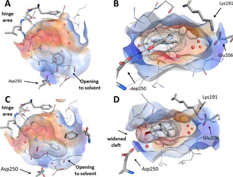
Fig 8. Comparison of co-crystal structures of 8g (PDB-ID: 6FT8; upper row) and 16 (PDB-ID: 6FT9; lower row) in complex with CLK1, respectively; red spheres: water molecules; black dashed lines: hydrogen bonds.
A: Top view of 8g. B: Side view of 8g. C: Top view of 16. D: Side view of 16. A, C: While the lactam motives of the ligands perform the canonical hydrogen bonds to the hinge region, the indole nitrogen atoms are connected to a conserved water molecule. B, D: An area near the Lys191 and Glu206 side chains, unoccupied by both 7g and 16, is filled by three water molecules. The opening to the entrance of the binding pocket is delimited by Asp250. Compared to the shape of the pocket with bound 8g (B), the entrance of the binding pocket is widened to accommodate the 2-bromo substituent of 16 (D).

